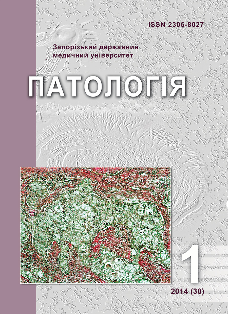
Ductular reaction in alcoholic steatohepatitis, nonalcoholic steatohepatitis and hepatitis C virus infection (immunohistochemical study)
DOI:
https://doi.org/10.14739/2310-1237.2014.1.25694Keywords:
Alagille syndrome, alcoholic steatohepatitis, nonalcoholic fatty liver disease, hepatitis CAbstract
Background and aim. Alcoholic steatohepatitis (ASH), nonalcoholic steatohepatitis (NASH) and hepatitis C virus infection (HCV) are the most frequent causes of liver cirrhosis in the Western world. The course of the disease depends on the balance in fibrogenesis, angiogenesis and regeneration. But if fibrosis was studied intensively during the last decades, mechanisms and signs of reparative regeneration in these diseases remain unknown. Ductular reaction is one of the morphologic signs of regeneration, but it was examined mostly in diseases associated with cholestasis. The aim of our work is to study ductular reaction associated with expression of immunohistochemical marker CK19 in alcoholic steatohepatitis, nonalcoholic steatohepatitis and hepatitis C virus infection.
Material and methods. 45 autopsies with diagnosed alcoholic steatohepatitis, nonalcoholic steatohepatitis and viral hepatitis C were enrolled in this study. Diagnosis of ASH was based on the data of alcohol abuse and morphologic signs of alcoholic disease – cardiomyopathy, chronic pancreatitis, alcoholic encephalopathy and typical liver changes. Viral genesis was proved by serological study (RNA HCV) and morphologic signs of HCV (METAVIR criteria). Diagnosis of NASH was verified by the features of metabolic syndrome and hepatic changes (Brunt criteria). The employed measures of reparative regeneration were CK19-positive cells with “biliary phenotype”, evaluated in the liver tissue (area of ductular reaction). Marker quantification was performed by assessing the ratio of stained tissue to the total area of the liver section using image analysis. Histotopographic features of ductular reaction and cellular phenotype were also studied. “STATISTICA FOR WINDOWS6.0” was used to analyze the data. The data were expressed as mean value ± standard deviation (M±s). Differences between groups were analyzed using ANOVA analysis (LSD test). P < 0,05 was considered significant.
Results. Evaluation of CK19-positive cells area in the liver section showed the highest degree in HCV group (10,17±0,23). Differences of this marker from the markers revealed in NASH (1,17±0,25) and ASH (1,99±0,4) were statistically significant. In addition to quantitative parameters localization of ductular reaction and cellular phenotype were analyzed. Diffuse spreading of the cells with “biliary phenotype” within the thickness of connective tissue septas was found in NASH and ASH. Most ductules were laid by homogenous cuboidal cells and contained distinct lumens. Probably these findings may be a result of biliary epithelial cell proliferation from the preexisting ductules. In HCV CK19-positive cells were revealed mostly in the site of marginal plate between the lobular parenchyma and connective tissue septa. In histologic examination cellular population with “biliary phenotype” was more heterogenous and some ductules had no lumens. Genesis of such lesions may be associated with activation and proliferation of immature precursors – intermediate hepato-biliary cells
Conclusions. Severe ductular reaction associated with marginal localization of CK19-positive cells between the parenchymal and connective tissue compartments is revealed in hepatitis C virus infection. In chronic steatohepatitis (alcoholic and nonalcoholic) diffuse spreading of the cells with “biliary phenotype” within the thickness of connective tissue septas is found. Quantitative and qualitative differences in morphologic signs of ductular reaction revealed in our examination confirm the variable role of new-formed ductules in the pathogenesis of different forms of chronic hepatitis which may be the aim for future researches.
References
Schuppan, D. & Afdhal, N. H (2008). Liver cirrhosis. Lancet, 371(8), 838–851. doi: 10.1016/S0140-6736(08)60383-9.
Williams, R. (2006) Global challenges in liver disease. Hepatology, (44), 521–-526. DOI: 10.1002/hep.21347.
Gural, A. L., Marievskij, V. F., Sergeeva, T. A. (2011). Harakteristika i tendencii razvitiya e`pidemicheskogo processa gepatita C v Ukraine [Characteristic and tendentions in the development of hepatitis C epidemic processes in Ukraine]. Profilaktychna medytsyna, (1), 9–17.
Riehle, R. J., Dan, Y. Y., Campbell, J. S. & Fausto, N. (2011). New concepts in liver regeneration. Journal of Gastroenterology and Hepatology, 26(1), 203–212.
Roskams, T. A., Theise, N. D. & Balabaud, C (2004). Nomenclature of the fi ner branches of the biliary tree: canals, ductules, and ductular reactions in human livers. Hepatology, (39), 1739–1745. DOI: 10.1002/hep.20130.
Zhang, L., Theise, N., Chua, M. & Reid, L.M. (2008). The stem cell niche of human livers: symmetry between development and regeneration. Hepatology, 48(5), 1598–1607.
Priester, S., Wise, C. & Glaser, S. S. (2010). Involvement of cholangiocyte proliferation in biliary fi brosis. World J.Gastroenterol. Pathophysiol., 1(2), 30–37. doi: 10.4291/wjgp.v1.i2.30.
Glaser, S. S., Gaudio, E. & Miller, T. (2009). Cholangiocyte proliferation and liver fi brosis. Expert Rev.Mol. Med., (30), 421–435.
Bedossa, P. & Poynard, T. (1996). An algorithm for grading activity in chronic hepatitis C. The French METAVIR Cooperative Study Group. Hepatology, (24), 289–293.
Brunt, E. M. & Tiniakos, D. G. (2010). Histopathology of nonalcoholic fatty liver disease. World Journal of Gastroenterology, 16(42), 5286–5296. doi: 10.3748/wjg.v16.i42.5286.
Dabbs, D. (2010). Diagnostic Immunohistochemistry. Theranostic and genomic application. New-York: Saunders Elsevier.
Popper, H., Kent, G. & Stein, R. (1957). Ductular reaction in the liver in hepatic injury. J Mt Sinai Hosp, (24), 551–556. 44
Downloads
How to Cite
Issue
Section
License
Authors retain copyright and grant the journal right of first publication with the work simultaneously licensed under a Creative Commons Attribution License that allows others to share the work with an acknowledgement of the work's authorship and initial publication in this journal.


