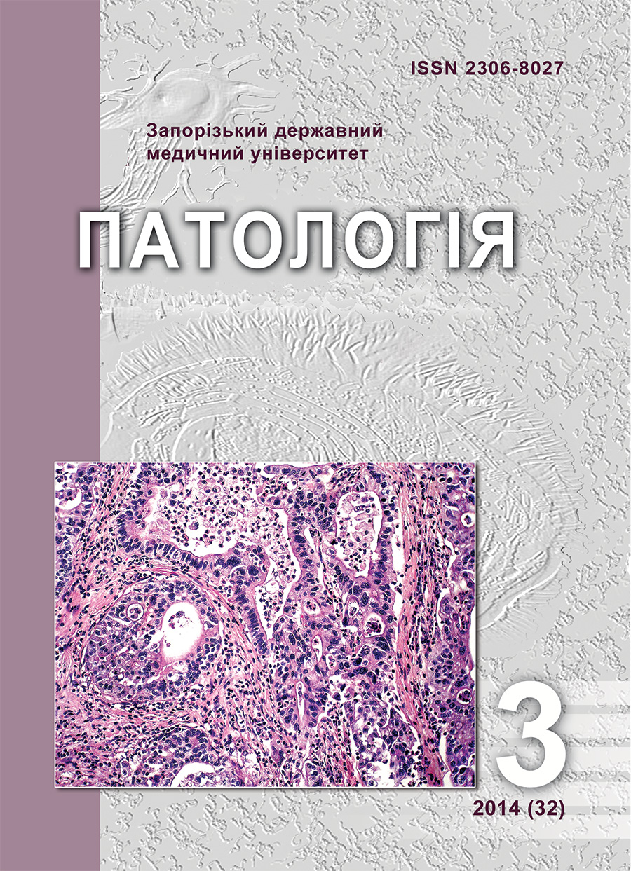
Morphological peculiarities of uterine tubes of fetuses from mothers with physiological pregnancy
DOI:
https://doi.org/10.14739/2310-1237.2014.3.36637Keywords:
Pregnancy, Fetus, Fallopian Tube, HistologyAbstract
Aim. In order to identify the structural features of the fallopian tubes in fetuses from women with physiological pregnancy at different stages of gestation, the structure of 47 antenatally dead fetuses were investigated.
Methods and results. Used such methods: anthropometric, organometric, histological, histochemical, morphometric and statistical. It has been established that in fetuses of 21st-28th weeks of gestation fallopian tubes are structurally completely formed, and at later dates the formation of their functional activity is noted.
Conclusion. This suggests that the effect of mother's pathology in early pregnancy leads to more serious violations of the structure of the fallopian tubes of the fetus. Described morphological features of the structure of the fallopian tubes of fetuses from mothers with physiological pregnancy according to gestational age can be used as a control group of observations in the study of structure of the organs of fetuses from mothers with complicated pregnancy.
References
Danilov, R. K. (2004). Gistologiya cheloveka v mul'timedia [Histology. Human multimedia]. Saint Petersnurg: ELBI-SPb. [in Russian].
Baranov, V. S. (2007) Novoe v prenatal'noj diagnostike i v
profilaktike nasledstvennykh i vrozhdennykh boleznej u ploda cheloveka [New in prenatal diagnosis and prevention of hereditary and congenital diseases in the human fetus]. Akusherstvo i ginekologiya, 5, 45–50. [in Russian].
Akhtemіichuk, Yu. T., & Piatnytska, T. (2010). Histotopohrafiia matkovykh trub u plodiv liudyny [Histotopohrafiya tubes in human fetuses]. Klinichna anatomiia ta operatyvna khirurhiia, 4, 50–54. [in Ukrainian].
Bashmakova, N. V., Kravchenko, E. N., & Lopushanskij, V. G. (2008). Rol' prognozirovaniya intranatal'nykh faktorov riska [Role predicting intrapartum risk factors]. Akusherstvo i ginekologiya, 3, 57–61. [in Russian].
Bagrіi, M. M., Demianchuk, M. V., Miller, I. V., et al. (2011). Histokhimichni metody doslidzhennia ekstratseliuliarnoho matryksu spoluchnoi tkanyny [Histochemical methods extracellular matrix of connective tissue]. Visnyk problem biolohii i medytsyny, 2, 248–251. [in Ukrainian].
Atramentova, L. A., & Utevskaya, O. M. (2008). Statisticheskie metody v biologii [Statistical methods in biology]. Gorlovka. [in Ukrainian].
Afanas`ev, Yu. I., & Yurina, N. L. (1989). Gistologija [Histology]. Moscow: Medicine. [in Russian].
Golubovskyi, I. A., Kostiuk, H. Ya., Korol, А. Р. (2006). Eksperymentalno-morfolohichne obgruntuvannia vidnovlennia prokhidnosti matkovykh trub [Experimental study morphological restoration of patency of the fallopian tubes]. Tavricheskij mediko-biologicheskij vestnik, 3, 50–52. [in Ukrainian].
Volkova, А., & Medvedieva, G. (2010) Faktory ryzyku nevynoshuvannia vahitnosti [Risk factors for miscarriage]. Proceedings of the 15th International Medical Congress of Students and Young Scientists, (р. 148), Ternopil. [in Ukrainian].
Zaporozhan, V. . (2006). Operatyvna hinekolohiia [Operative gynecology]. Odesа: University Press. [in Ukrainian].
Downloads
How to Cite
Issue
Section
License
Authors who publish with this journal agree to the following terms:- Authors retain copyright and grant the journal right of first publication with the work simultaneously licensed under a Creative Commons Attribution License that allows others to share the work with an acknowledgement of the work's authorship and initial publication in this journal.

- Authors are able to enter into separate, additional contractual arrangements for the non-exclusive distribution of the journal's published version of the work (e.g., post it to an institutional repository or publish it in a book), with an acknowledgement of its initial publication in this journal.
- Authors are permitted and encouraged to post their work online (e.g., in institutional repositories or on their website) prior to and during the submission process, as it can lead to productive exchanges, as well as earlier and greater citation of published work (SeeThe Effect of Open Access).

