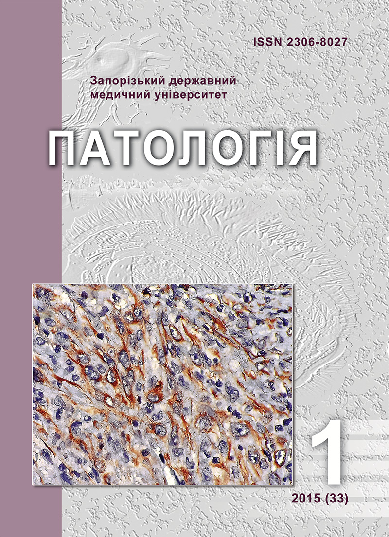
Pan cytokeratin expression by meningiomas of the brain
DOI:
https://doi.org/10.14739/2310-1237.2015.1.42828Keywords:
Meningioma, Histogenesis, CytokeratinAbstract
Meningiomas of the brain are the second most common tumor of the CNS in adults, accounting about 30% of all primary tumors.
Aim. In order to determine the presence and to assess the expression level of cytokeratin (CK) in benign and malignant brain meningiomas tissue samples were obtained during neurosurgical operations from 30 patients.
Methods and results. Tissue samples were studied immunohistochemically by using monoclonal antibodies Mo a-Hu Cytokeratine (PanCK), Clone AE1/AE3 («DAKO», Denmark). It was established that anaplastic meningiomas expressed CK in 80% of cases. Among benign meningiomas positive expression was found in 40% of fibroblastic subtypes.
References
Louis, D. N., Ohgaki, H., Wiestler, O. D., & Cavenee, W. K. (Eds.). (2007). World Health Organization Classification of Tumours of the Central Nervous System. Lyon: IAR.
Miller, R. Jr., DeCandio, M. L., Dixon-Mah, Y., Giglio, P., Vandergrift, W. A. 3rd, Banik, N. L., et al. (2014). Molecular Targets and Treatment of Meningioma. Journal of Neurology and Neurosurgery, 1(1).
Pekmezci, M., & Perry, A. (2013). Neuropathology of brain metastases. Surgical Neurology International, 4, S245–55, doi: 10.4103/2152-7806.111302.
Bekyashev, A. Kh. (2011). [Pathogenesis of meningiomas (a review of literature)]. Opukholi holovy i shei, 4, 26–40. [in Russian].
Abdelzaher, E., & Abdallah, D. M. (2014). Expression of mesothelioma-related markers in meningiomas: an immunohistochemical study. Biomed Research International. doi: 10.1155/2014/968794.
Babichenko, I. I., & Kovyazin, V. A. (2008). Novyie metody immunogistokhimicheskoj diagnostiki opukholevogo rosta [New methods of immunohistochemical diagnosis of tumor growth]. Moskow: RUDN. [in Russian].
Liu, Y., Sturgis, C. D., Bunker, M., Saad, R. S., Tung, M., Raab, S. S., & Silverman, J. F. (2004). Expression of cytokeratin by malignant meningiomas: diagnostic pitfall of cytokeratin to separate malignant meningiomas from metastatic carcinoma. Modern Pathology, 17(9), 1129–1133. doi:10.1038/modpathol.3800162.
Theaker, J. M., Gatter, K. C., Esiri, M. M., & Fleming, K. A. (1986). Epithelial membrane antigen and cytokeratin expression by meningiomas: an immunohistological study. Journal of clinical pathology, 39(4), 435–439. doi: 10.1136/jcp.39.4.435.
Taraszewska, A., & Matyja, E. (2014). Secretory meningiomas: immunohistochemical pattern of lectin and ultrastructure of pseudopsammoma bodies. Folia Neuropathologica, 52(2), 141–150.
Chung, B. M., Rotty, J. D., & Coulombe, P. A. (2013). Networking galore: Intermediate filaments and cell migration. Current opinion in cell biology, 25(5), 600–612. doi: 10.1016/j.ceb.2013.06.008
Downloads
How to Cite
Issue
Section
License
Authors who publish with this journal agree to the following terms:- Authors retain copyright and grant the journal right of first publication with the work simultaneously licensed under a Creative Commons Attribution License that allows others to share the work with an acknowledgement of the work's authorship and initial publication in this journal.

- Authors are able to enter into separate, additional contractual arrangements for the non-exclusive distribution of the journal's published version of the work (e.g., post it to an institutional repository or publish it in a book), with an acknowledgement of its initial publication in this journal.
- Authors are permitted and encouraged to post their work online (e.g., in institutional repositories or on their website) prior to and during the submission process, as it can lead to productive exchanges, as well as earlier and greater citation of published work (SeeThe Effect of Open Access).

